
Department of Radiology
- The Department of Radiology is the primary department responsible for diagnostic imaging at the University's medical center, which includes 865 hospital beds and multiple large outpatient facilities in Ann Arbor and nearby communities.
- The combination of a large tertiary referral center and community practices, provides trainees the opportunity to learn from an outstanding breadth of pathologies and clinical presentations.
- Over 700,000 examinations are performed annually.
U-M Rogel Cancer Center and Breast Care Center
- The U-M Rogel Cancer Center is a designated National Comprehensive Cancer Network (NCCN) site.
- Within the Cancer Center, the Breast Care Center provides consultation and management for approximately 700 new cancer patients per year.
- Every week breast imagers participate in the Breast Care Center’s Multidisciplinary Tumor Board and help optimize individualized treatment plans for patients.
- Breast imagers enjoy a high level of camaraderie with clinical colleagues.
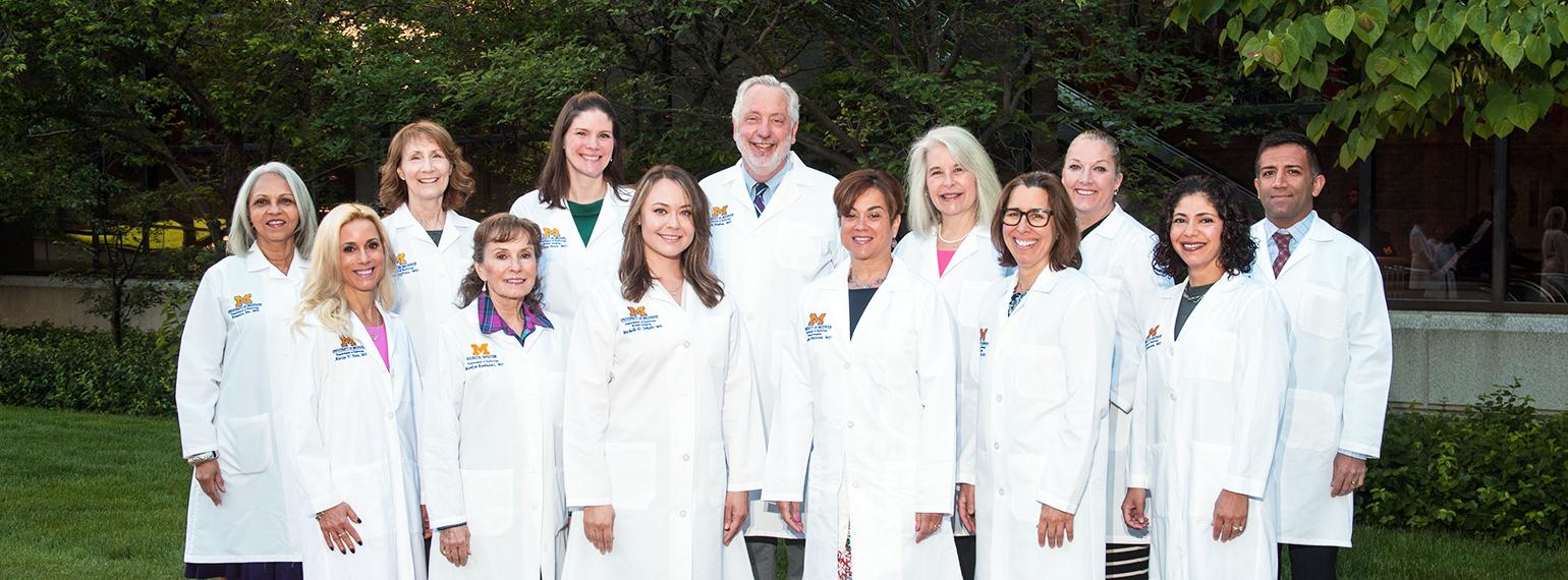
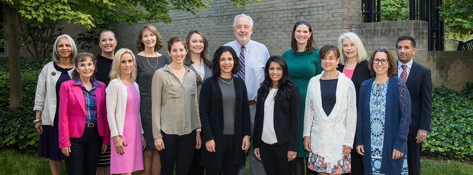
Breast Imaging Division
- 15 MQSA-certified breast radiologists provide diagnostic imaging services at three sites, two within Ann Arbor and one in Brighton, Michigan. Offline screening mammograms are performed at four additional imaging sites in nearby communities.
- Greater than 50,000 breast imaging studies are interpreted annually.
- Trainees and faculty routinely perform diagnostic ultrasounds, although dedicated breast sonographers are also an integral part of our breast imaging team.
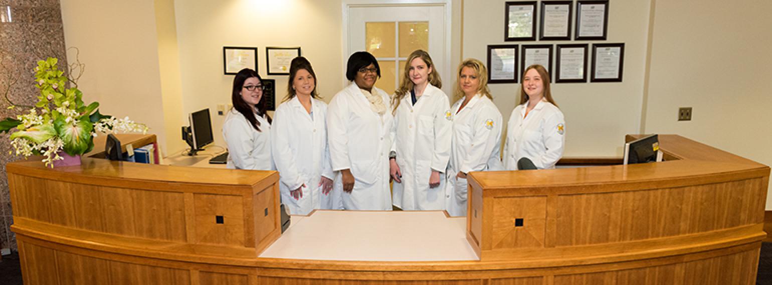
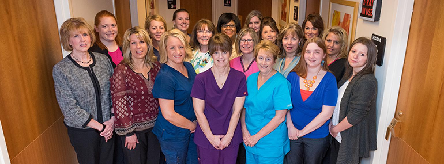
- Our technologists, nurses, and clerical staff are critical to our success in delivering optimal, efficient patient care.
The Breast Imaging Fellowship Experience
We offer a post-residency breast imaging fellowship that is designed to provide individuals an opportunity for specialized training and academic development. Fellows will actively participate in all functions of the Breast Imaging Division and will assume significant responsibility for its daily operation under faculty supervision. Research and teaching opportunities supplement the clinical curriculum.
We strive to develop relationships with our fellows that endure beyond their training. Our graduates have been successful in both academic and private practice.
Curriculum
Experience
The fellowship consists of in-depth training in all aspects of breast imaging. On average, fellows complete more than 2,500 breast imaging studies during their 12-month fellowship.
- Screening and diagnostic mammography
- Digital breast tomosynthesis
- Breast MRI
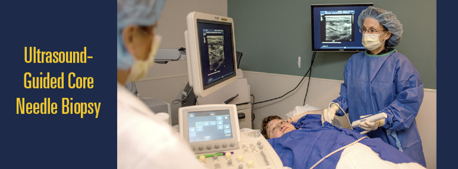
- Performing breast ultrasound
- Proficiency in performing all image-guided procedures
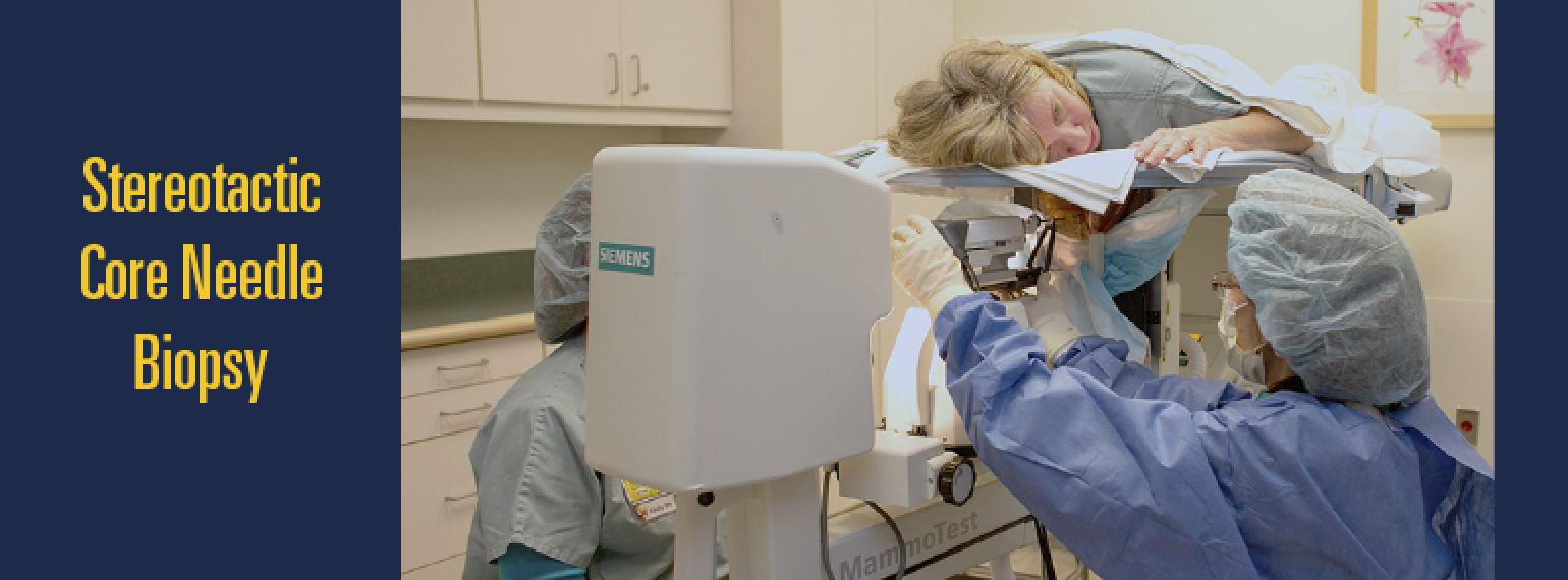
Annually, over 600 preoperative localizations (including Magseed), 900 core biopsies, and 300 aspiration procedures are performed by the division.
Education
- Weekly Multidisciplinary Tumor Board. This is a great way to see numerous breast cancers and learn about their management.
- Didactic and case-based lectures are given by faculty
- Weekly breast imaging conference rotates between radiology-pathology correlation, journal club, and interesting case conference.
- Interdisciplinary experiences in breast surgery, pathology, and radiation oncology
- Familiarity with MQSA requirements and FDA inspections
The fellow is provided informal and formal opportunities to teach trainees and colleagues.
Research
The Department of Radiology’s strong research programs in ultrasound, MRI, computer-aided diagnosis, and physics offer a multimodality approach to the study and diagnosis of breast disease.
- Fellows complete one research project during the year. They often present at a national radiology meeting.
- Non-clinical time is given to fellows involved in research investigations.
- Historically, fellow research has been published in leading journals such as Radiology or American Journal of Roentgenology.
Fellow Publications
Below is a list of fellow publications:
- Reducing Number of Patient Visits and Time to Biopsy After Suspicious Breast MRI. Rahman WT, Moorman SEH, Neal CH, Hall RA, Cheasick HL, Arnold D, Pujara AC. J Am Coll Radiol. 2022 Feb;19(2 Pt A):251-253. doi: 10.1016/j.jacr.2021.10.023. Epub 2022 Jan 15. PMID: 35041854
- Annual Screening Mammography Associated With Lower Stage Breast Cancer Compared With Biennial Screening. Moorman SEH, Pujara AC, Sakala MD, Neal CH, Maturen KE, Swartz L, Egloff H, Helvie MA. AJR Am J Roentgenol. 2021 Jul;217(1):40-47. doi: 10.2214/AJR.20.23467. Epub 2021 May 5. PMID: 33955776
- Neal CH, Sakala M, Houck G, Noroozian M, Kazerooni EA, Davenport MS. Improving Breast MR Wait Times: A Model for Transitioning Newly Implemented Diagnostic Imaging Procedures into Routine Clinical Operation. JACR 2018; 15(6):859-864.
- Savage J, Jeffries DO, Noroozian M, Sabel M, Jorns J, Helvie MA. Pleomorphic Lobular Carcinoma In Situ: Imaging Features, Upgrade Rate, and Clinical Outcomes. AJR 2018;211:1-6.
- Neal CH, Rahman WT, Joe AI, Pinsky RW, Noroozian M, Helvie MA. Harms of Restrictive Risk-Based Mammographic Breast Cancer Screening. American Journal of Roentgenology. 2018;210:1–7.
- Rahman WT, Neal CH, Brown RJK. Nuclear Medicine Imaging and Management of Breast Lesions. Educational Exhibit at the Society of Nuclear Medicine and Molecular Imaging Annual Meeting. June 2018. Philadelphia, Pennsylvania.
- Bosma MS, Neal CH, Klein KA, Noroozian M, Patterson SK, Helvie MA. Does Direct Radiologist-Patient Verbal Communication Affect Follow-Up Compliance of Probably Benign Assessments? J Am Coll Radiol 2016; 13(3): 279–285. PM: 26777739
- Bosma MS, Morden KL, Klein KA, Neal CH, Knoepp US, Patterson SK. Breast imaging after dark: patient outcomes following evaluation for breast abscess in the emergency department after hours. Emerg Radiol 2016;23(1):29-33. PM: 26433916
- Coletti MC, Joe AI, Jeffries DO, Wey E. Localized Amyloidosis of the Breast Mimicking Breast Cancer: Radiologic-Pathologic Correlation. Med Res Archives 2015; (3):1-6.
- Noroozian M, Stein LF, Gaetke-Udager K, Helvie MA. Long-term clinical outcomes in women with breast pain in the absence of additional clinical findings: Mammography remains indicated. Breast Cancer Res Treat 2015;149(2):417-424. PM: 25556516
- McLaughlin CT, Neal CH, Helvie MA. Is the upgrade rate of atypical ductal hyperplasia diagnosed by core needle biopsy of calcifications different for digital and film-screen mammography? AJR Am J Roentgenol 2014;203(4):917-922. PM: 25247961
- Yilmaz ZN, Neal CH, Noroozian M, Klein KA, Sundaram B, Kazerooni EA, Stojanovska J. Imaging of Breast Cancer–Related Changes After Nonsurgical Therapy. AJR Am J Roentgenol 2014; 202(3):675-683. PM: 24555607
- Neal CH, Yilmaz ZN, Noroozian M, Klein KA, Sundaram B, Kazerooni EA, Stojanovska J. Imaging of breast cancer-related changes after surgical therapy. AJR Am J Roentgenol 2014;202(2):262-272. PM: 24450664
- Neal CH, Coletti MC, Joe A, Jeffries DO, Helvie MA. Does digital mammography increase detection of high-risk breast lesions presenting as calcifications? AJR Am J Roentgenol 2013;201(4):1148-1154. PM: 24147490
- Neal CH, Daly CP, Nees AV, Helvie MA. Can preoperative axillary US help exclude N2 and N3 metastatic breast cancer? Radiology 2010;257(2):335-341. PM: 20807849
- Daly CP, Bailey JE, Klein KA, Helvie MA. Complicated breast cysts on sonography: is aspiration necessary to exclude malignancy? Acad Radiol 2008;15(5):610-617. PM: 18423318
- Martin KE, Helvie MA, Zhou C, Roubidoux MA, Bailey JE, Paramagul C, Blane CE, Klein KA, Sonnad SS, Chan HP. Mammographic density measured with quantitative computer-aided method: comparison with radiologists' estimates and BI-RADS categories. Radiology 2006;240(3):656-665. PM: 16857974
- Foster MC, Helvie MA, Gregory NE, Rebner M, Nees AV, Paramagul C. Lobular carcinoma in situ or atypical lobular hyperplasia at core-needle biopsy: Is excisional biopsy necessary? Radiology 2004;231(3):813-819. PM: 15105449
- Kaiser JS, Helvie MA, Blacklaw RL, Roubidoux MA. Palpable breast thickening: Role of mammography and US in cancer detection. Radiology 2002;223(3):839-844. PM: 12034957
- Guest AR, Helvie MA, Chan HP, Hadjiiski LM, Bailey JE, Roubidoux MA. Adverse effects of increased body weight on quantitative measures of mammographic image quality. AJR Am J Roentgenol 2000;175(3):805-810. PM: 10954471
Equipment
State-of-the-art equipment includes:
- Fifteen mammography units
- Six digital breast tomosynthesis units
- Six ultrasound units with high resolution transducers and Doppler capabilities
- A prone digital biopsy table with tomosynthesis capability and vacuum assisted core biopsy capabilities
- A 3 Tesla Philips magnet with dedicated 16-channel breast coil and biopsy capability
Living in Ann Arbor
- This University town of approximately 120,000, is a comfortable place to live and work for families and young adults alike. It is frequently ranked among the top best cities in which to live.
- The University and city provide a wealth of art, cultural, and educational experiences throughout the year.
- It is a major scene of college sports, boasting the largest football stadium in the world.
- There is an excellent bus system and the city is bike-friendly.
- The Huron River and many city parks provide opportunities for outdoor relaxation and recreational activities.
- Michigan, otherwise known as the Great Lakes state, has innumerable beautiful destinations in its lower and upper peninsulas, for those who love adventure, summer and winter sports.
- An international airport, Detroit Metropolitan (DTW), is less than 30 minutes driving distance from the city.
To learn more, go to Life at Michigan.
Apply
For the 2025-2026 academic year, interested applicants may electronically submit their completed application (please see application file at the bottom of this page; alternatively, the SBI Universal Application will also be accepted) to [email protected] November 1 - December 31, 2023.
We will participate in the NRMP Match. Virtual interviews will be held in February 2024. If COVID safety guidelines allow, we may consider accommodating individual requests for on-site visits.
Questions regarding our program may be directed to:
Carol McLaughlin, M.D.
c/o Carol Kruise
Michigan Medicine
Department of Radiology
A. Alfred Taubman Health Care Center
1500 E. Medical Center Drive, TC2910
Ann Arbor, MI 48109-5326
Ph. 734-936-4367
Fax. 734-936-9723
[email protected]
Downloads
Fellowship Director
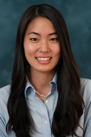
Carol McLaughlin, MD
Associate Fellowship Director