
Positron Emission Tomography (PET) Imaging
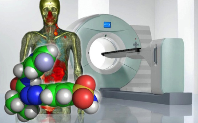
Positron emission tomography (PET) imaging is a nuclear medicine imaging technique that can be used to non-invasively elucidate biochemical processes, diagnose disease and track the impact of experimental drugs in clinical trials – all in living human subjects.
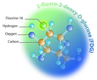
Patients receive an injection of radiopharmaceutical, a drug labeled with a positron emitting radionuclide (e.g. carbon-11, fluorine-18), followed by a PET scan. The most common radiopharmaceutical is [18F]fluorodeoxyglucose (FDG), a radioactive sugar molecule that can be used to identify tumors and diagnose cancer:
During the PET scan procedure, the PET-CT scanner detects pairs of gamma rays emitted, indirectly, by the radiopharmaceutical.
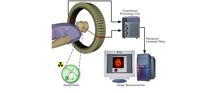
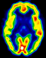

Images of radiopharmaceutical concentration in 3-dimensional or 4-dimensional space within the body are then reconstructed by computer analysis to provide physicians with an image of, for example, the patient’s brain or the whole body. In modern scanners, this reconstruction is typically accomplished with the aid of a CT scan performed on the patient during the same procedure.
The radiochemistry group actively participates in industry-supported collaborative research projects in radiochemistry and preclinical studies of new drugs and radiopharmaceuticals, and provides routine delivery of radiopharmaceuticals for clinical trials with partners in the pharmaceutical and radiopharmaceutical drug industry.