
Purpose
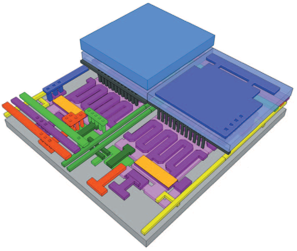
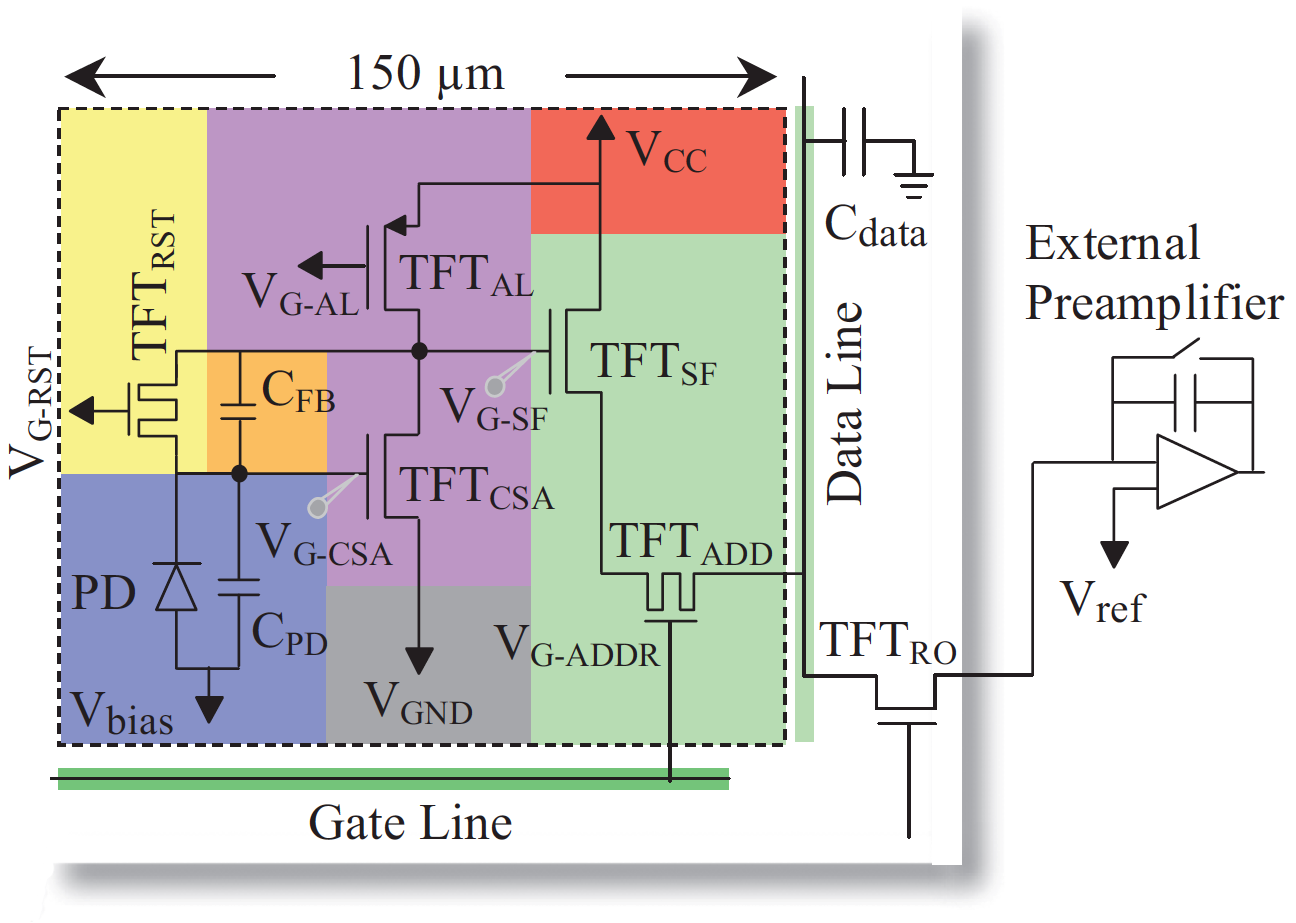
To develop imager backplanes consisting of pixel circuits incorporating amplifiers based on poly-Si thin-film transistors (TFTs). The goal is to overcome the performance limitations of active matrix, flat-panel imagers (AMFPIs) at low x-ray dose while preserving the advantages of AMFPI technology – thereby improving image quality in applications such as fluoroscopy and CBCT. The incorporation of an amplifier in the pixel circuit (referred to as the active pixel concept) allows the imaging system to maintain favorably high signal-to-noise performance over a greater range of imaging conditions. The adjacent drawing is a schematic 3D rendering of a 5-TFT pixel design whose circuit is shown in the accompanying diagram – with both illustrations color-coded to match corresponding circuit elements. The circuits are based on poly-Si – a semiconductor material that facilitates fabrication of very large area, monolithic circuits which exhibit good radiation damage resistance.
Background
Following extensive research by our group and others, active matrix, flat-panel imagers (AMFPIs) have become ubiquitous for projection and tomographic medical imaging. Their uses range from the treatment of cancer with high-energy radiation beams to diagnosis of disease and interventional procedures. However, AMFPI technology suffers from significant limitations that improvement to the signal-to-noise properties can overcome.
Impact
The improved performance expected from the additional gain in signal provided by an in-pixel amplifier should enhance image quality under conditions of low radiation dose to the patient. This improvement, along with other new capabilities offered by poly-Si circuits such as extremely high frame rates, could facilitate advanced applications such as very low dose fluoroscopy, breast and chest tomosynthesis, dual-energy imaging and cone-beam computed-tomography for angiographic, breast, radiotherapy and other procedures.
Approach
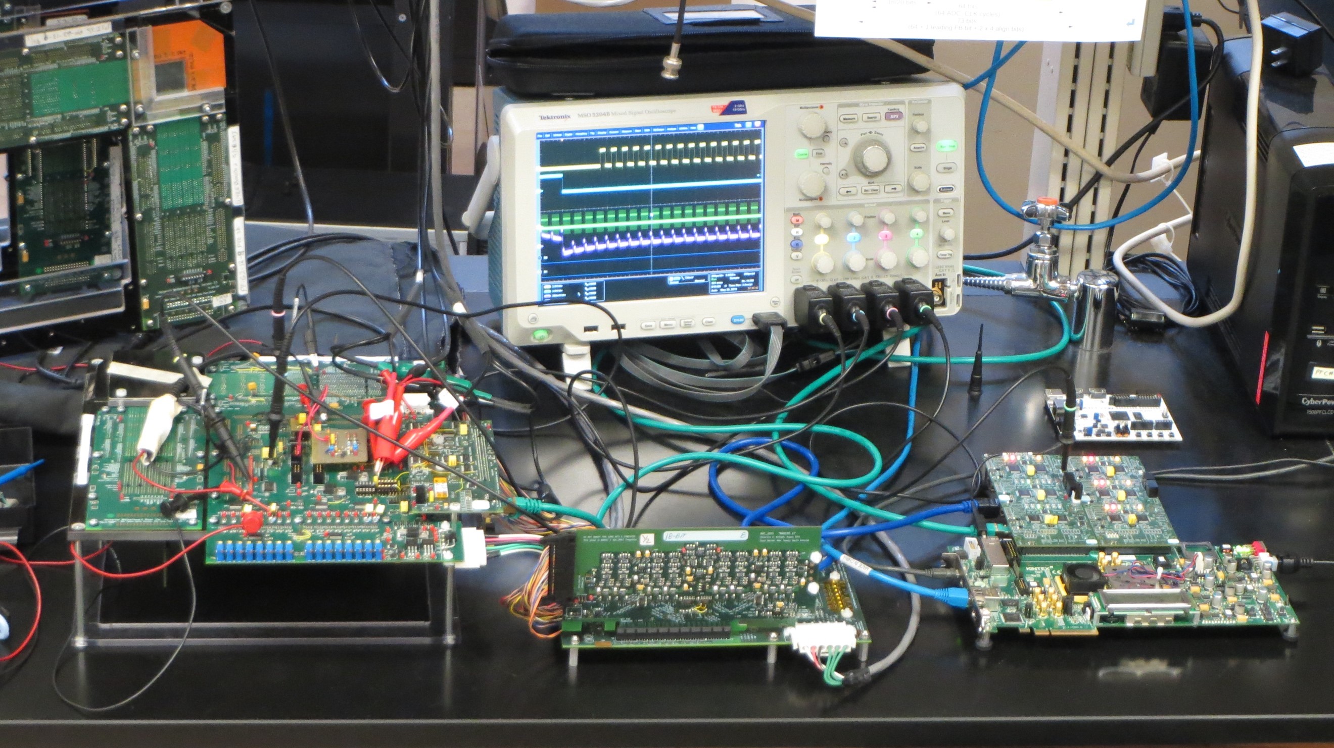
In partnership with industrial collaborators, our group designs, fabricates and evaluates sophisticated prototype imagers in which the pixel circuits incorporate out-of-plane and three-dimensional circuit architectures comprising in-pixel amplification structures. A small area, prototype backplane (representing an order-of-magnitude increase in circuit complexity compared to that of contemporary AMFPI pixel designs) along with a custom-made, electronic data acquisition system is shown in the adjacent photo.
Project Funding
This research was supported by the National Institutes of Health / National Institute of Biomedical Imaging and Bioengineering as well as by our department.
Recent Publications
Koniczek et al., SPIE Vol. 10573, Physics of Medical Imaging, 2018: 105730L-1 to 105730L-8.
Koniczek et al., Med. Phys. 44(7), 3491-3503, 2017.
Antonuk et al., SPIE Vol. 8668, Physics of Medical Imaging, 2013: 86680A-1 to 86680A-9.
Koniczek et al., SPIE Vol. 7961, Physics of Medical Imaging, 2011: 79610P-1 to 79610P-10.
El-Mohri et al., Med. Phys. 36(7), 3340-3355, 2009. PMCID: PMC2805355.
Antonuk et al., Med. Phys. 36(7), 3322-3339, 2009. PMCID: PMC2807886.
Antonuk et al., SPIE Medical Imaging, 725814, 2009.
Antonuk et al., Mater. Res. Soc. Symp. Proc. 1066, 1066-A19-03, 2008. PMCID: PMC2941962.
Antonuk et al., SPIE Medical Imaging, 69130I, 2008.
Li et al., J. Appl. Phys. 99, 064501-1 to 064501-7, 2006.
Antonuk et al., SPIE Vol. 5745, Physics of Medical Imaging, 2005: 18-31.