
Purpose
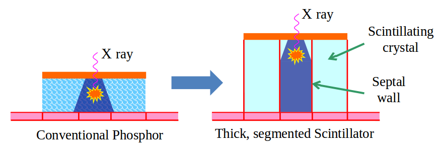
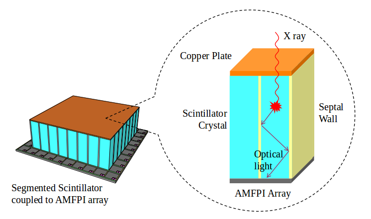
To develop new forms of x-ray scintillators for imaging the megavoltage beam used to treat cancer in external-beam radiation therapy. The goal is to improve visualization of soft tissues in tomographic x-ray images acquired at low, clinically practical x-ray doses. Our lab is developing detectors based on thick segmented crystalline scintillators to replace the relatively thin phosphor scintillators presently used in active matrix, flat-panel imagers (AMFPIs) designed for radiotherapy. Such devices can also be adapted to the needs of various forms of non-destructive testing.
Background
Following pioneering research conducted by our lab, AMFPIs have become the gold standard for creating projection images of the patient using the high-energy, x-ray treatment beams employed in radiation therapy. However, the low x-ray detection efficiencies (~2%) of present megavoltage imagers strongly limit their clinical utility.
Impact
The significant improvements in x-ray detection efficiency sought in our research will make it possible to achieve soft-tissue contrast, even at the lowest doses delivered by radiotherapy treatment machines. Moreover, such improvements will make it possible to acquire tomographic imaging information (requiring ~100 projection images) at the same dose presently required for a single projection image. Such improvements are expected to significantly assist the basic medical objective of maximizing the dose to the tumor while minimizing dose to surrounding normal tissue.
Approach
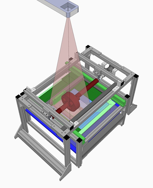
In partnership with industrial and national lab collaborators, we design, fabricate and/or evaluate prototype segmented scintillating converters providing x-ray detection efficiencies ranging from ~7% to 50%. The performance of these prototypes is extensively investigated and can include demonstrations of computed tomography using experimental set-ups such as that depicted in the adjacent illustration.
Project Funding
This research has been funded by the National Institutes of Health / National Cancer Institute, and by Alamos National Lab as well as by our department.
Publications
Liu et al., Med. Phys. 42(4), 2072-2084, 2015
Liu et al., Med. Phys. 41(6), 061916, 2014. PMCID: PMC4039737
El-Mohri et al., Phys. Med. Biol. 59(4), 797-818, 2014. PMCID: PMC4061715
Liu et al. Phys. Med. Biol. 57(16), 5343-5358, 2012. PMCID: PMC3429122
El-Mohri et al., Phys. Med. Biol. 56(6), 1509-1527, 2011. PMCID: PMC3062516
Wang et al., Phys. Med. Biol. 55(13), 3659-3673, 2010. PMCID: PMC2909124
Wang et al., Med. Phys. 36(12), 5707-5718, 2009. PMCID: PMC2797046
Wang et al., Med. Phys. 36(7), 3227-3238, 2009. PMCID: PMC2805354
Wang et al., Med. Phys. 35(1), 145-158, 2008. PMCID: PMC2920060