Middle and Inner Ear Delivery Systems
The Miller Laboratory has extensively used the micro-cannula and Alzet osmotic pump to deliver agents into the middle ear or directly into scala tympani of the guinea pig (Brown et al). Several changes to the delivery system have allowed for longer, more reliable infusion times (Prieskorn & Miller).
- Using the same micro-cannula, we have developed a method of bolus delivery of agents in the guinea pig scala tympani.
- To expand research potential, we developed a micro-cannula for use in mouse or rat, which also allowed access to additional sites in the guinea pig inner ear (scala media, scala vestibuli).
- Another system designed and used successfully in our lab for years is an injection port system mounted on the guinea pig head with a cannula accessing the ear. This device is utilized with agents that would be affected by body temperature or when larger volumes are needed for delivery into the middle ear of the guinea pig.
- Another development is a cannula/electrode device which allows simultaneous delivery of agents to the scala tympani along with electrical stimulation.
Electrode Stimulation System
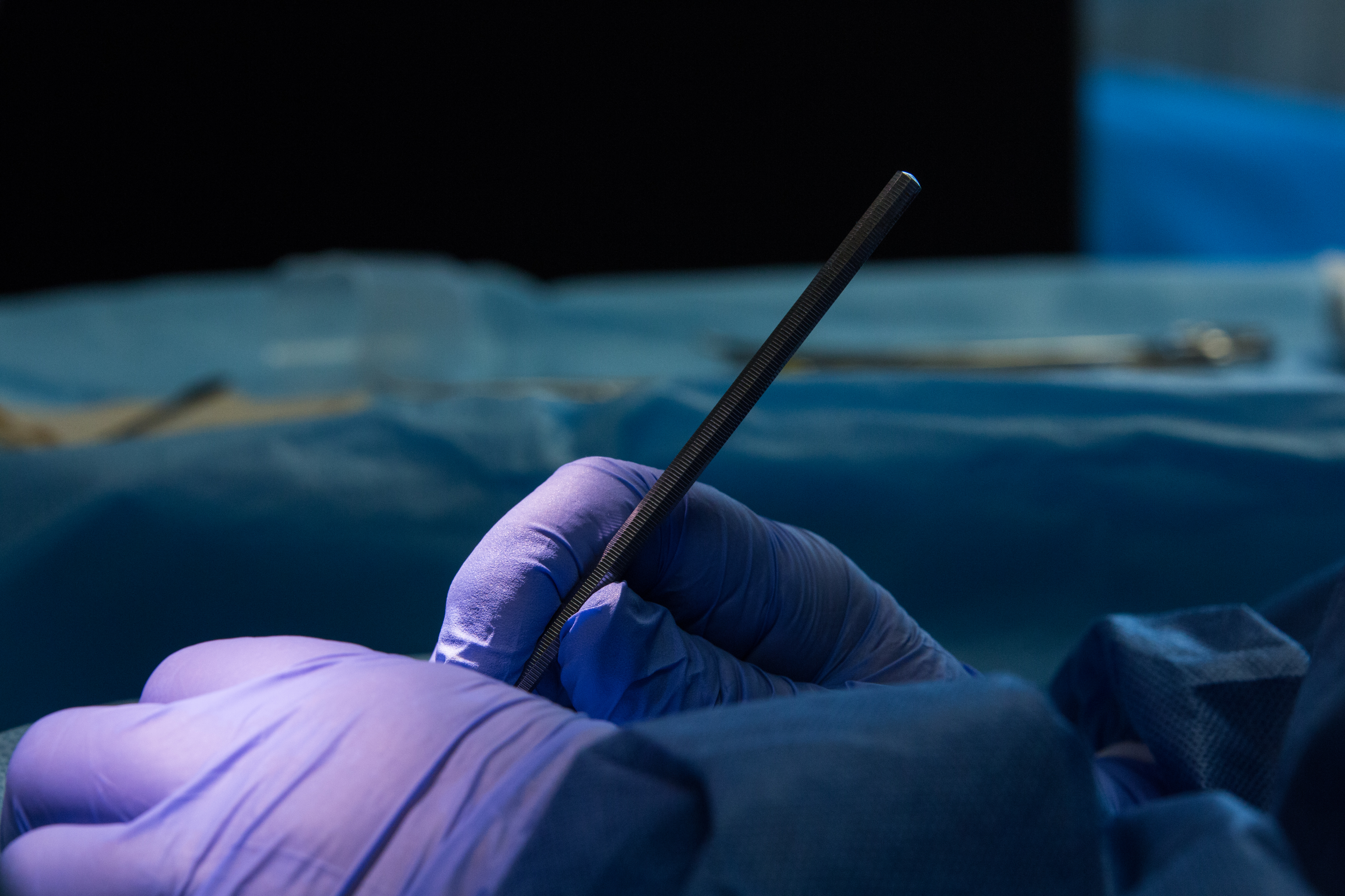
- We make electrodes from platinum/iridium wire for implantation into scala tympani or on the round window of the guinea pig. The electrodes are attached to a percutaneous connector that is mounted on the dorsal surface of the head.
- A stimulation device, developed in-house by Mr. Chris Ellinger, allows us to plug into the electrode and provide stimulation to the inner ear. We can control the amplitude and duty cycle.
- Another development is a cannula/electrode device allowing simultaneous delivery of agents to the scala tympani along with electrical stimulation.
Cytocochleograms
A computer program was developed in-house by Dr. David B. Moody to facilitate tabular and graphic presentation of cochlear sensory cell loss in guinea pig and mouse models. Microscope slide mounts of inner ear tissue are assessed for sensory cell loss, and missing cell counts are entered into the program. A graph (cytocochleogram) is generated depicting cell loss as a function of distance along the basilar membrane of the inner ear. This depiction is useful in various studies involving, e.g., noise exposure and drug infusions.
