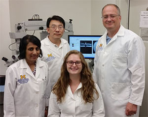
Managers Naheed Khan and Cheng-mao Lin, staff member Sarah Sheskey, and director David Antonetti
- Director: David Antonetti, PhD
Phone: 734-232-8230
Email: [email protected] - Manager: Cheng-mao Lin, PhD
Phone: 734-232-8220
Email: [email protected] - Manager (ERG): Naheed Khan, PhD
Phone: 734-764-4163
Email: [email protected] - Staff: Sarah Sheskey, BS
Phone: 734-998-5896
Email: [email protected]
Submit a work order to the Functional Assessment Core
The Functional Assessment Core provides training and support to help investigators perform non-lethal measures of ocular structure and function. Currently, the Core provides expertise in OCT, OptoMotry, ERG, fundus imaging, and pupillometry. Please call or email us to discuss your project needs and how the Functional Assessment Core can help advance your research.
Equipment
The equipment can be reserved for use after training. Reserve Equipment here. A computer workstation is also available for data processing.
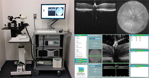
Bioptigen Envisu Preclinical Spectral Domain Optical Coherence Imaging System
- Real-time non-invasive imaging of the microstructure of retinal tissue in small laboratory animals (rats and mice)
- Acquire, process, display and save depth-resolved images of ocular microstructure
- Repeat imaging for longitudinal studies
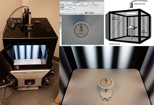
Cerebral Mechanics OptoMotry System
- Real time behavioral testing of optokinetic response for small laboratory animals (rats and mice)
- Visual acuity and contrast sensitivity testing
- Repeat testing for longitudinal studies
Phoenix Micron III Retinal Imaging System
- Bright field and fluorescent retinal imaging for small laboratory animals (rats and mice)
- High resolution images in live animals
- Repeat imaging for longitudinal studies

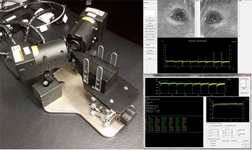
NeurOptics A-2000 Small Animal Pupillometer System
- Tracking and recording pupil size change in small laboratory animals (rats and mice) in real time
- Binocular camera system with two independently controlled visible light sources in 4 colors and infrared illumination for various stimulation modes
- Repeat imaging for longitudinal studies
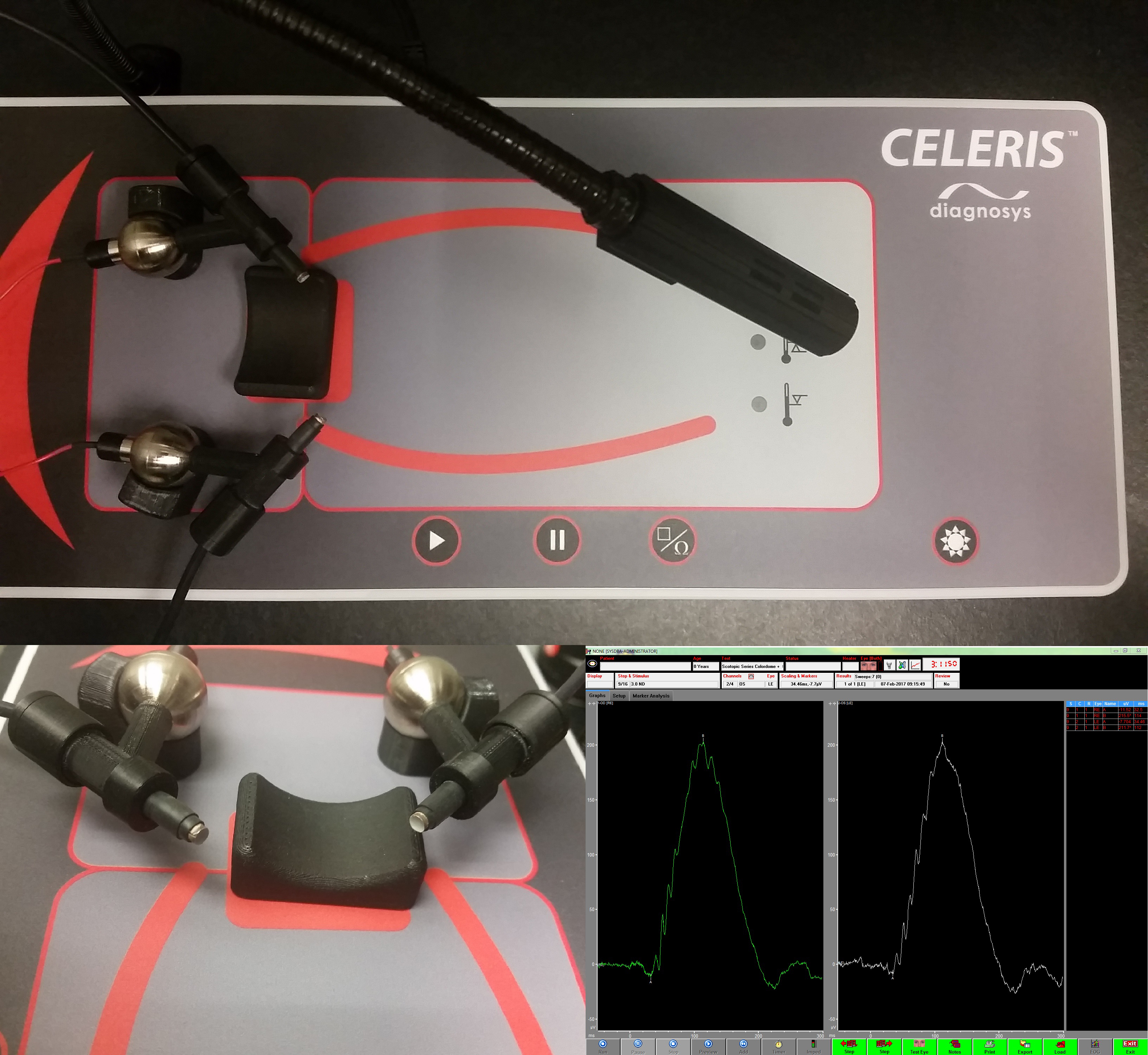
Diagnosys Celeris Electrophysiology System
- Perform full-field dark-adapted and light-adapted ERGs
- Ability to stimulate both eyes independently
- Ability to run tests with one eye as a reference, or test both eyes simultaneously
- Capability to perform pattern ERGs
- Repeat testing for longitudinal studies
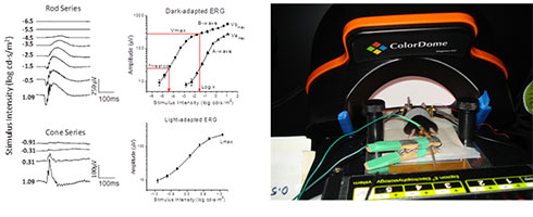
Diagnosys Espion E2 Electrophysiology System
- The Espion E2 electrophysiology system comprises hardware, software and stimulators to perform full-field dark-adapted and light-adapted electroretinograms (ERGs) and other electrophysiological tests.
- Custom protocols include single flash rod response, single flash cone response, flicker response, scotopic threshold response, ON-OFF response, oscillatory potentials, photopic negative response.
- Stimulator
- LEDs, produce any color flash and background.
- Xenon flash for brighter flashes, maximum intensity is 3000 cd.s/m2.
- The recording set up consists of a grounded and shielded Faraday cage and temperature controlled warming bed.
- Capability to perform pattern ERGs