

Clinical Contacts
Administrative Contact
Biography
Larry E Antonuk, PhD is the principal investigator of the Flat Panel Imager Group and a Professor in the Physics Division within the Department of Radiation Oncology of the School of Medicine, located on the Medical Campus of The University of Michigan in Ann Arbor. He is also affiliated with the Biomedical Engineering Department within the College of Engineering at the University.
Following his PhD and postdoctoral nuclear physics studies in Canada, Switzerland, and France, Dr. Antonuk came to the University of Michigan in 1987. There he founded the Flat-panel Imager Lab, and his early research focused on the design, fabrication and empirical characterization of active matrix, flat-panel imagers (AMFPIs) – a technology based on thin-film electronics that has become ubiquitous in medicine. His lab focuses on the development of technologies offering order-of-magnitude improvement in performance compared to existing projection and volumetric x-ray imagers for medical applications such as radiotherapy, radiography, fluoroscopy, breast imaging, cardiac and interventional imaging, as well as destructive and non-destructive testing.
Areas of Interest
- Our research concentrates on development of the two major components of a flat-panel x-ray imager: the large-area, monolithic, solid-state backplane which takes the form of a two-dimensional matrix of imaging pixels whose thin-film circuitry is a focus of the research; and the converter positioned over the backplane where incident x-rays interact and which consists of either a scintillator or a photoconductor.
- The following monolithic, large-area solid-state x-ray imager technologies under development in our lab offer prospects for significant improvements in performance, or new capabilities, compared to existing projection and volumetric x-ray technologies.
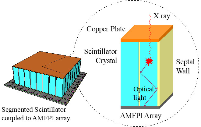
Technology 1: Thick, segmented scintillator converters – for external-beam, radiotherapy imaging and non-destructive testing.
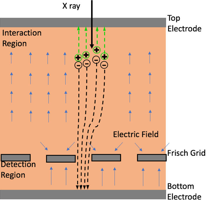
Technology 4: Photon-counting imaging backplanes (consisting of arrays that comprise highly-complex pixel circuits based on poly-crystalline silicon) which measure the spectral information of each interacting x-ray photon – for Cone Beam Computed Tomography in radiotherapy (kV-CBCT) and Breast Computed Tomography (BCT).
- The research methodology is based on iterative design, fabrication and performance evaluation of experimental imager configurations. These configurations comprise prototype arrays or converters (typically fabricated by industrial partners) that are packaged into the mechanical and electronic sub-systems (created by our group) that are required to fully test them. This methodology has been found to be a cost-effective means of developing advanced imager designs.
- Computer simulations of radiation transport and optical transport in x-ray converters, as well as the signal and noise properties of advanced backplane circuits, are an essential element of our research. Such simulations are computationally expensive and, for that reason, are carried out using a custom-designed, in-house computer cluster – one of the numerous state-of-the-art tools utilized in the lab.
Clinical Interests
- Investigating x-ray imaging technologies for applications such as radiotherapy, radiography, fluoroscopy and breast imaging, as well as cardiac and interventional imaging.
Honors & Awards
2011 Inaugural inductee, League of Research Excellence of the University of Michigan Medical School
2006 Dean’s Award for Innovation from the University of Michigan Medical School
2004 Fellow, The American Association of Physicists in Medicine
Credentials
PhD, University of Alberta, 1980
Grants
- Subcontract No.370629, (PI Antonuk), 4/2016-10/2018, Los Alamos National Laboratories. Development and Analysis of Scintillator Array.
- R01-EB0022028, (PI Antonuk), 9/2016-5/2020, National Institute of Biomedical Imaging and Bioengineering. Development of a High Sensitivity, X-ray Detector Technology Based on Polycrystalline Mercuric Iodide for Volumetric Breast Imaging.
Published Articles or Reviews
Selected from 137 publications
- Liang AK, Koniczek M, Antonuk LE, El-Mohri Y, Zhao Q, Street RA, Lu JP. Performance of In-Pixel Circuits for Photon Counting Arrays (PCAs) Based on Polycrystalline Silicon TFTs. Phys Med Biol. 61(5):1968-1985, 2016.
- Liu L, Antonuk LE, El-Mohri Y, Zhao Q, Jiang H. Theoretical investigation of the design and performance of a dual energy (kV and MV) radiotherapy imager. Med Phys. 42(4):2072-2084, 2015.
- Jiang H, Zhao Q, Antonuk LE, El-Mohri Y, Gupta T. Development of active matrix flat panel imagers incorporating thin layers of polycrystalline HgI2 for mammographic x-ray imaging. Phys Med Biol. 58(3):703-714, 2013.
- El-Mohri Y, Antonuk LE, Koniczek M, Zhao Q, Li Y, Street RA, Lu JP. Active pixel imagers incorporating pixel-level amplifiers based on polycrystalline-silicon thin-film transistors. Med Phys. 36(7):3340-3355, 2009.
- Antonuk LE, Zhao Q, El-Mohri Y, Du H, Wang Y, Street RA, Ho J, Weisfield R, Yao W. An investigation of signal performance enhancements achieved through innovative pixel design across several generations of indirect detection, active matrix, flat-panel arrays. Med Phys. 36(7):3322-3339, 2009.